Otology/Neurotology
(110) Computed Tomography as an Adjunct for Cadaveric Temporal Bone Drilling
Monday, October 2, 2023
2:45 PM - 3:45 PM East Coast USA Time

Has Audio

Silas Chao, MD
Resident
Tripler Army Medical Center
AIEA, Hawaii, United States- BC
Brian S. Chen, MD
Neurotologist
Tripler Army Medical Center
Glendale, HI, USA
Presenting Author(s)
Senior Author(s)
Disclosure(s):
Silas Chao, MD: No relevant relationships to disclose.
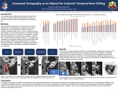
Introduction: Determine the educational value of the novel use of serial Computed Tomography (CT) scans of cadaveric temporal bones during temporal bone dissection lab.
Methods: This is a Prospective cohort study over two years (2020-2021). Six otolaryngology residents were surveyed prospectively on the usefulness of serial CT scans of cadaveric temporal bones prior to and after temporal bone dissection. CT scans were performed on cadaveric temporal bones a total three times: first prior to any dissection, second after initial dissection of a canal wall up procedure, and third after a canal wall down procedure. CT images were reviewed by the operative resident and the staff otologist after each acquisition. Pre and post drilling Likert (1-7) surveys rating the confidence in identifying 14 radiologic landmarks on CT imaging as well as 20 surgical landmarks intraoperatively were collected. Final practicum grading of a simple mastoidectomy with facial recess was assessed objectively and scores were compared to prior year’s scores without use of CT as an adjunct to the dissection.
Results: There was overall increased confidence of the residents in both identifying radiographic structures on CT and intraoperatively. There was higher pre-dissection confidence in radiologic landmarks than in surgical landmarks. 0/14 radiologic and 6/20 surgical landmarks had statistically significant improvement (p < 0.05) in post-drilling confidence compared to pre-drilling confidence. There was a greater improvement of confidence levels in junior residents compared to senior residents. Final practicum scores showed a consistent positive trend of improved scores as PGY group increased over the study period.
Conclusions: The role of serial CT images as an adjunct to temporal bone dissection is useful in familiarizing the novice surgical resident to the complex anatomy and correlating the surgical and radiologic anatomy of relevant landmarks. Interval scans also provided feedback on the extent, thoroughness, and safety of temporal bone dissection.
